Also referred to as epidermoid or dermoid cysts.
These are slow growing benign lesions that result from an error of cell migration during embryonic development.
These lesions develop when cells in the developing embryo that were destined for the skin, hair or nail tissue become entrapped in the developing brain or spinal cord.
These trapped cells ultimately produce what is called an inclusion cyst and the cyst contents can vary depending on the exact cell type that was trapped
Tumor | Location | Contents |
Epidermoid | Cerebellopontine angle, Sylvian fissure, skull base | Epidermal debris Mother of pearl |
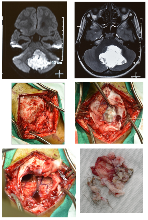
Dermoid | Mid line and skull base | Hair and sebaceous material apart from epidermal debris |
Symptoms
The symptoms of an intracranial epidermoid and dermoid are caused by the pressure the growing tumor exerts on surrounding brain.
They can be headaches, nausea, seizures, vision loss, facial pain, weakness or numbness in the limbs or face, and gradual changes in mood or personality.
If the cyst ruptures and spill its contents, it can cause repeated bouts of severe meningitis, with symptoms including fever, headache, and neck stiffness.
Treatment
Surgical excision-
The primary treatment for symptomatic dermoid or epidermoid cysts is surgical removal.
Goals of surgical removal are to remove the cyst contents but to also remove the cyst lining if possible.
Chemotherapy- No role
Radiation therapy- No much role
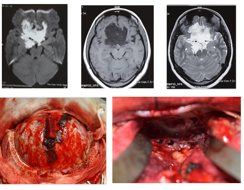
Subfrontal epidermoid:
45 YRS/ M
Headache
Visual blurring
Cerebellar dermoid:
12Yrs/ M
Headache, Vomiting, Imbalance